")

")
")
")
Canine Ulna Model,Study Model Dog Ulna Model,Medical Anatomical Dog Ulna Model,Animal Skeleton Bone Anatomy Model,For Orthopaedic Surgery Demonstration Practice China Manufacturer.
Introduction: The Canine Ulna Model emerges as an invaluable resource in the realm of veterinary education and clinical practice, offering an immersive learning experience and realistic surgical simulations. With its tailored customization options, this model caters to a diverse array of educational objectives, making it an indispensable asset in both classroom settings and clinical environments.
Versatility: As a Study Model Dog Ulna Model, this anatomical representation transcends conventional boundaries, serving as a versatile tool for various educational and practical purposes. Its adaptability allows seamless integration into clinical skills labs, surgical workshops, and educational demonstrations, providing students and practitioners alike with an immersive learning platform.
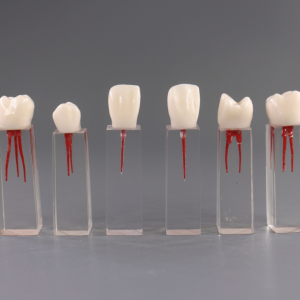
Realism: Anatomical accuracy lies at the core of the Canine Ulna Model, meticulously replicating the intricate details and structural nuances of a canine ulna bone. From the curvature to the articulating surfaces, each component mirrors the complexities of real anatomy, ensuring a lifelike representation essential for effective learning and surgical practice.
Quality: Crafted from high-quality materials, this model exemplifies durability and reliability. Constructed with medical-grade PU, it withstands the rigors of repeated use while retaining its integrity. Corrosion-resistant and lightweight, it offers unparalleled longevity without compromising on performance, guaranteeing years of reliable service.
Customization: One of the standout features of the Canine Ulna Model is its customizable nature, allowing for tailored simulations to meet specific educational objectives. From common clinical scenarios to rare pathological conditions, instructors can design case-specific models to enhance learning outcomes and provide a more comprehensive understanding of veterinary orthopedics.
Functionality: Primarily designed for orthopedic surgery simulation, the Canine Ulna Model facilitates hands-on learning in a controlled environment. Its 1:1 life-size scale and realistic anatomical features enable students to practice surgical techniques, refine their skills, and prepare for real-world clinical scenarios. Additionally, its versatility extends to classroom demonstrations, fostering interactive learning experiences.
Conclusion: In summary, the Canine Ulna Model emerges as an indispensable tool in veterinary education and clinical practice, offering unparalleled realism, durability, and versatility. With its tailored customization options and immersive learning features, it elevates the standard of anatomical study aids and surgical simulations, empowering students and practitioners to excel in their respective fields.